All about morphea: a specific form of localized scleroderma
Morphea is a rare skin disease, belonging to the family of localized scleroderma. It is characterized by the appearance of thickened skin lesions, sometimes discolored, which indicate skin inflammation followed by skin fibrosis. Despite its localized nature, this condition requires particular vigilance to prevent complications and preserve the quality of life of affected individuals.

What is morphea and how to recognize it?
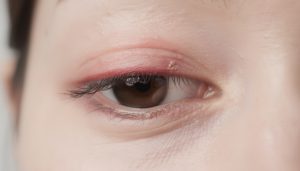
Morphea differs from systemic scleroderma in that it generally remains confined to the skin layer, without affecting internal organs. The skin plaques, sometimes oval and showing a thick hardening, may have a lilac border, while the center changes color from red to white or yellow. This evolution reflects the inflammatory process followed by fibrosis that alters the texture and appearance of the skin.
There are several forms of the disease, ranging from limited plaque morphea to linear morphea, often seen in children, with elongated bands on the face or limbs. This more severe variant can affect underlying tissues, muscles, or even bones, sometimes leading to deformities.
Main causes and risk factors of morphea
The causes of morphea remain partially unclear, but current research highlights several leads. The onset seems linked to an autoimmune response where the immune system overreacts and mistakenly attacks skin tissue. This causes skin inflammation followed by excessive collagen production, the protein responsible for skin fibrosis.
In addition, environmental factors such as repeated skin trauma, exposure to chemical agents, and possibly viral infections play a role, although direct evidence is limited. A genetic predisposition is often suspected, especially in individuals with a family history of autoimmune diseases.
Moreover, lifestyle elements such as chronic stress and smoking could worsen symptoms or contribute to the onset of lesions.

Factors that increase the risk of developing morphea:
- Age: more commonly diagnosed in children and young adults.
- Sex: women are more frequently affected.
- Family history: presence of autoimmune diseases in the family.
- Environmental exposure: contact with toxins or irritating chemical agents.
- Coexisting autoimmune diseases: lupus, rheumatoid arthritis.
Recognizing the signs of morphea for early diagnosis
Morphea symptoms are not uniform and may include:
- Appearance of thickened and discolored plaques, often oval.
- Itching or localized discomfort at the lesions.
- In some cases, limited mobility when a joint is near an affected area.
- Hair loss in cases where lesions involve the scalp.
Lesions may evolve slowly or rapidly, sometimes spreading, which requires urgent medical consultation in case of increased redness, pain, or signs of infection.
Morphea diagnosis: methods and precautions
To establish a morphea diagnosis, the dermatologist first performs a thorough clinical examination, assessing the appearance and location of lesions. The medical history, family history, and symptom evaluation are crucial.
Additional tests, such as skin biopsy, aim to confirm the inflammatory and fibrosing nature of the lesions. Blood tests help exclude other associated autoimmune diseases. Sometimes, medical imaging assesses deeper tissue involvement.
Morphea treatment options to limit progression and relieve symptoms
Management of morphea is adapted to the form and severity of the lesions. Treatments aim to reduce inflammation, limit fibrosis, and improve skin comfort.
- Topical corticosteroids: applied to plaques to soothe inflammation.
- UV phototherapy: used to reduce skin thickening in some cases.
- Immunosuppressive drugs: such as methotrexate for extensive or severe forms.
- Physiotherapy: to prevent or treat joint contractures related to lesions.
- Complementary approaches: stress management, anti-inflammatory diet, gentle skin care.
Regular medical monitoring is essential to adjust therapy and prevent long-term complications.